Breast MRI
Magnetic resonance imaging is one of the most precise and informative diagnostic methods. With the help of magnetic field and radio waves it produces high quality whole-body and organ cross-sectional images in three planes in one sitting; without harming the body.
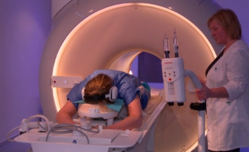
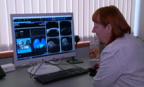
Breast MRI examinations are carried out at our new ARS Diagnostic clinic with the first fully digitalised magnetic resonance equipment in Latvia – Philips Ingenia 3,0T.
 |
 |
Magnetic resonance imaging in breast diagnostics is used:
- if ultrasonography or mammography does not give precise enough information but cancer cells have been detected in axillary lymph nodes or if there is an increased risk of developing cancer;
- when breast cancer has been diagnosed – to determine the course of the disease, to specify its location and distribution in order to ascertain whether there is another outbreak and plan further treatment tactics and/or extent of the operation. During the examination both breasts are examined;
- in order to establish the number and size of tumors in a breast that contains multiple tumors. It is also used to clarify ambiguous formations found with previous methods;
- to evaluate the efficiency of chemotherapy;
- to ascertain that previously treated tumor has not returned;
- if it is necessary to accurately assess breast structure and/or implant condition after plastic surgery.
Recommendation:
To avoid changes in breasts caused by hormonal fluctuation, patients are recommended to undergo their MRI examination during the 2nd week of the menstrual cycle (between days 5-12). This guarantees significantly more accurate results.
Contrast agent use during breast examinations
A contrast agent is used in an MRI when it is necessary to evaluate breast structure and different formations. The contrast agent is administered into a vein. In order to ensure maximum safety, the patient must inform their radiologist of any kidney diseases, allergic reactions and medications used at the time of the examination.
Contrast agents are not necessary when only assessing breast implant condition.
Newest technologies have a very important role in breast examinations.
In comparison to other available MRI machines in Latvia the Philips Ingenia 3,0T, located in the “ARS” Diagnostic clinic, is the only fully digitalised 3 Tesla strength equipment capable of providing a fast, very high resolution examination.
The examination is harmless and painless.
MRI should not be performed if a patient has:
- pacemakers,
- nerve stimulators,
- cochlear implants,
- heart valve prostheses or various other metal implants specified as unsuitable for MRI.
If a patient suffers from claustrophobia – fear from enclosed spaces, the attending physician can administer calming medication prior the procedure.
How to prepare for MRI examination:
- To maximize the use of MRI diagnostic capacity, patients need to take with them all previous ultrasound and mammography results and statements of previous surgery, manipulations or other treatments, extracts from the hospital, etc., relating to the disease in question.
- The patient’s blood must be analysed to determine their creatinine levels, the result must be taken to the MRI examination.
- Women are advised to avoid using make up that could contain tiny metal particles (mascara, eyeshadow), as in the effect of magnetic field they can contaminate the equipment and reduce image quality.
MRI examinations are performed by:
- Diagnostic radiologist – Dr Biruta Rasma VAGULE
- Diagnostic radiologist – Dr Ilze ENĢELE
Note:
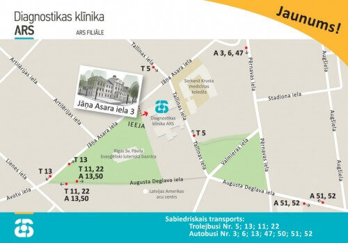
Magnetic resonance examinations are carried out at the Medical company ARS branch – ARS Diagnostic clinic,
- Address – Jāņa Asara ielā 3, Rīgā
Magnetic Resonance Imaging (3 tesla):
- registration for the survey: +371 672 01 088
- information about the results of the survey: +371 66 929 760

