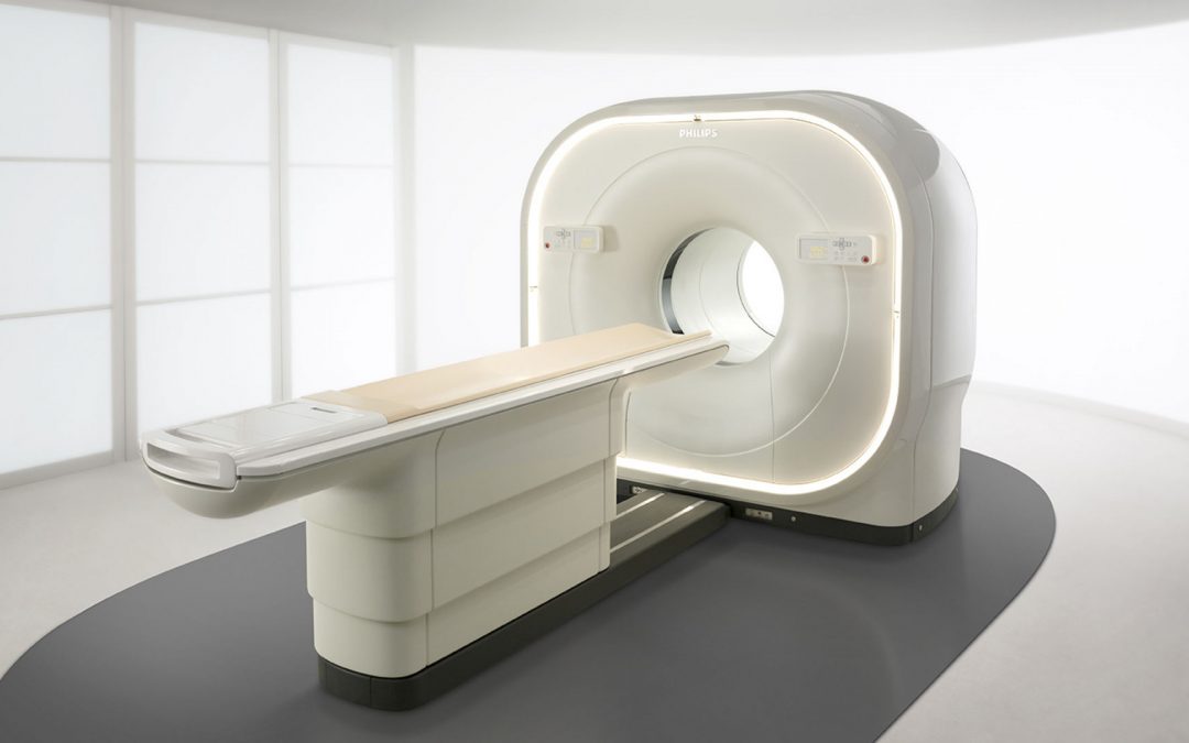
Medicine Centre ARS launches examinations by using innovative digital PET/CT (positron emission tomography) equipment.
Digital PET/CT equipment offers significant advantages:
- Unlike the analogue equipment a digital detector can register each unique photon, which appears in tissues by decaying radiopharmaceutical preparations, thereby considerably enhancing the equipment capacity to visualize tiny foci, which, likely, would not be seen on the analogue equipment.
- Digital PET/CT examinations enable to analyze the structure of the damage in more details, thereby enabling to differentiate the tumours better.
- New technologies enable to shorten the examination time considerably and reduce the total radiation load received.
Why PET/CT examination?
PET/CT is a combined examination, which consists of two examinations, where as a result three informative images are obtained.
- PET (positron emission tomography) equipment visualizes such areas in the body, where as a result of increased rate of metabolic activity a radiopharmaceutical preparation, which contains glucose analogues, or matches other specific cell receptors, is attracted more. An increased rate of glucose metabolism and increased expression of some receptors is intrinsic to tumour cells.
- CT (computer assisted tomography) examination renders the image of the patient anatomic structure, where it is possible see the organs, take measurements and determine the size of formations
The obtained PET and CT images are digitally layered on top of each other, thereby obtaining information of both anatomic structures and tissue physiological characteristics – metabolic activity.
When PET/CT examination is assigned?
- Where a patient has been diagnosed with a malignant tumour and such tumour has been proven, PET/CT examination is highly valuable to establish whether the disease has not spread further over the body, to identify precisely the stage of the disease and select the treatment tactics.
- To assess the treatment effectiveness, for instance, after surgery or chemotherapy, also after the cancer recuperation, to exclude relapse of the disease.
- Examination sometimes helps identify the primary tumour, detect its location and differentiate between benign and malignant changes.
Examination procedure
Each patient is previously assessed whether in a particular case the PET/CT method is appropriate.
To perform PET examination a radiopharmaceutical preparation is administered into the patient’s vein. This preparation at various intensities accumulates in the body tissues and, once decayed, causes the photon flow, which is registered by a PET detector. Just before PET examination the patient undergoes CT with or without contrast.
PET/CT examines the body from the base of the skull to the midthigh, if necessary, examines completely the whole body.
The examination is completely safe and painless. It takes about 20-40 minutes, but it is necessary to take into account yet another 2 hours to spend in the clinic to make preparations before the examination.
After the examination the patient can go home. The radioactive substance at the end of the examination decays to the level harmless to the environment and to the wider public.
After the examination the radiologists analyse the images obtained, reply to the questions put by the consulting physician in the referral.
The reply can be received personally or electronically within 5 working days.
How to get appointment to PET/CT examination?
The examination is performed with the referral from a physician only.
PET/CT examination is performed in the following cases:
- breast cancer – to exclude distant metastasis at stage III;
- bronchus, lung cancer – at stage I-III;
- colorectal cancer – to asses previously diagnosed distant metastasis for the potentially operable patients;
- melanoma – at stage III or IV;
- urological malignancy;
- gynaecological malignancy;
- gastrointestinal stomal tumours;
- for children, if such decision is taken by the paediatric haematology /oncology medical consilium;
- by a decision of a haematology medical consilium – in cases of extramedullary dissemination of lymphoid tissue malignant tumours.
| PET/CT examinations are assessed by a radiologist diagnostician MD Marika KALNIŅA – one of the most experienced specialists in Latvia in the PET/CT method. |
 |
ARS Diagnostic Clinic
J. Asara iela 3, Rīga
PET/CT coordinator: (+371) 26 533 446
E-mail: pet@arsmed.lv
www.ars-med.lv