
Breast ultrasound (US) refers to one of non-invasive tissue examination methods. Ultrasound is often required to complement mammography, but it cannot substitute mammography with tomosynthesis either in terms of sensitivity or specificity.
The key objective of breast ultrasound is:
- Distinguish dense formation from hollow or fluid-containing formation;
- In case of dense breast, if palpatory finding is uncertain;
- To confirm and more carefully examine a damage, which is seen on mammographic image only in one projection;
- Assisting in diagnostic manipulations, such as aspiration biopsy;
- During post-surgical period up to 6 months, also after surgery on breast augmentation and implant insertion, specifically in cases of their damaging.
Ultrasound examination is also recommended to:
- Young women;
- Pregnant women;
- Women during breastfeeding (lactation) period;
- men.
US as the primary breast examination method is recommended to:
- young and nulliparous women under 30 y/o.
- women under 30 y/o with a small breast
- pregnant women
- women with breast implants
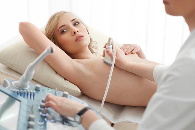
Examination procedure
During examination the patient is lying flat on back, the breast is placed firmly on the chest front wall, thereby reducing density of the breast itself. Examination is performed with a slight pressure to reduce density of beneath tissues, by applying high frequencies 7.5 – 11.0 MHz, a small (4cm) linear probe. Examination depth is 4 cm and less.
Breast is examined simultaneously with auxiliary vaults, evaluating the skin, subcutaneous, glandular tissues (internal mammary structures such as segments and milk ducts), adipose tissues, which are located under the breast and chest wall (including muscles and ribs), detailed assessment of nipples.
Key advantages of breast ultrasound:
- examination using the ultrasound method is radiation free;
- this procedure is not unpleasant to the patient;
- examination using the ultrasound method is easy to perform and location of the formation is easy to describe;
- breast is comparatively transparent and well examinable throughout;
- suspicious formations can be detected at the initial stage;
- examination is easy to repeat.
Basic breast examination methods are ultrasound and mammography. Each of these methods provides its own information of mammary glands, but in combination both methods complement each other, thereby enabling to obtain optimal information of mammary glands.
Breast ultrasound examination is performed by:
| Medical clinic “Medicīnas centrs ARS” Skolas Street 5, Riga Phone: +371 67 201 007 |
ARS Diagnostic Clinic J. Asara Street 3, Riga Phone: +371 66 929 750 |