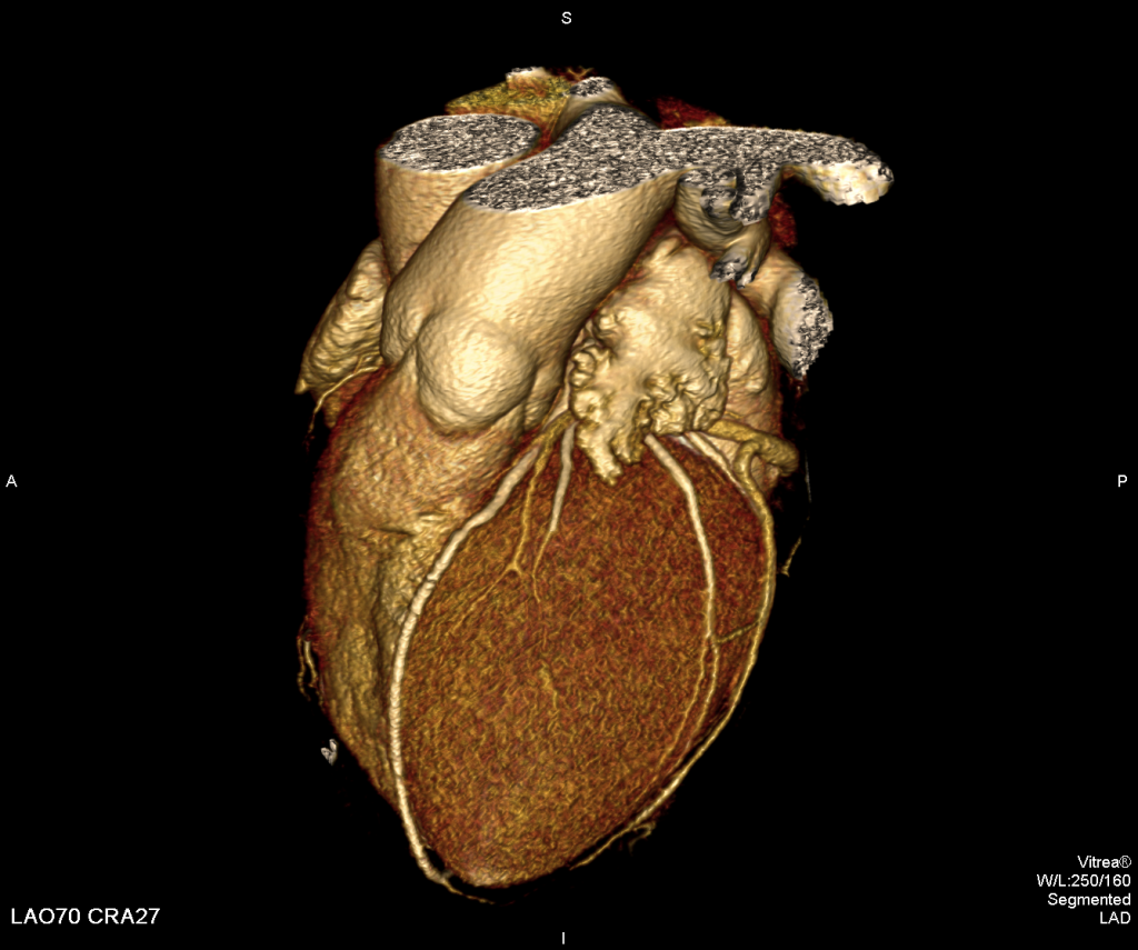
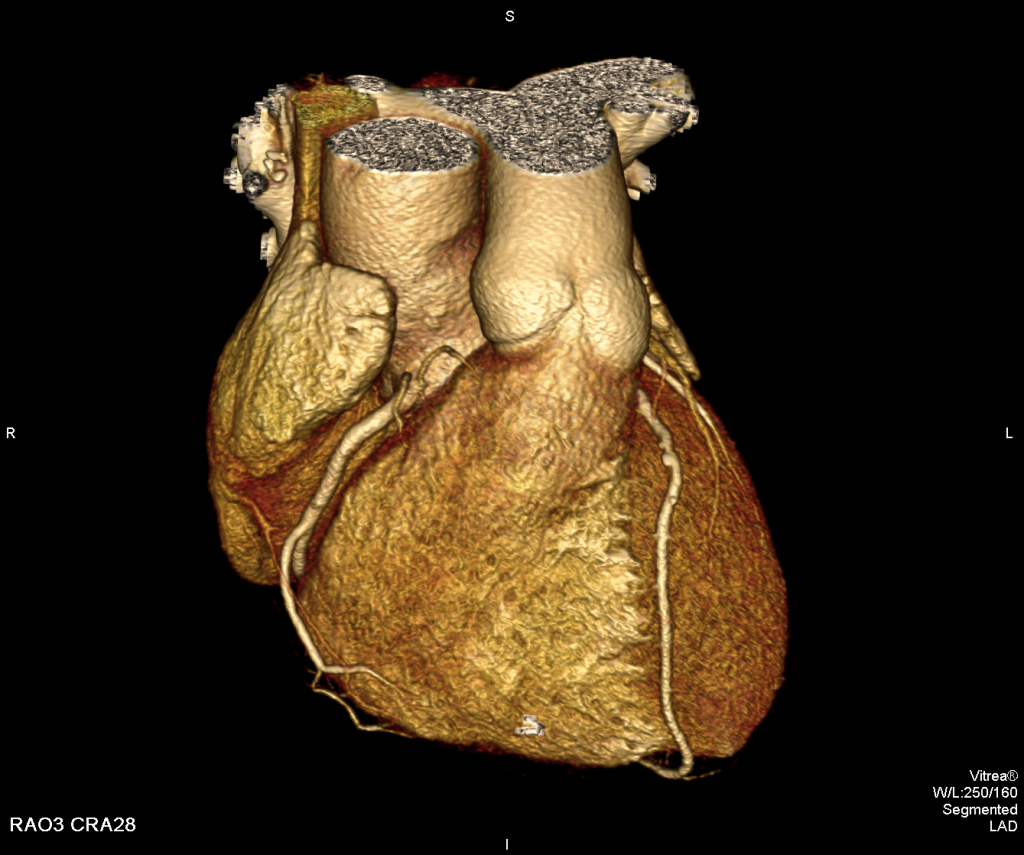
CT coronary angiography is a non-invasive method to identify the condition of the heart (coronary) blood vessels. This method allows assessing the risk of heart diseases, making prognosis without exposing patients to an unpleasant, invasive procedure – catheterization of blood vessels. The examination is a painless procedure, which can be performed at an outpatient setting. This service at our Clinic is paid up within the framework of the state programme, if the patient has a GP referral.
CT coronary angiography indications:
- Screening – early detection of atherosclerosis in the coronary blood vessels and screening of patients for conventional angiography. The risk group encompasses patients with the increased stress, cholesterol level and overweight. Attention should be paid to men over 40 y/o, women over 55 y/o.
- In case of atypical chest pain syndrome.
- For the control of stents and coronary artery bypass and condition evaluation.
- After unsuccessful invasive coronary angiography.
The objective of CT coronary angiography
is to determine the condition of coronary arteries, but additionally it is possible to obtain information about:
- Cardiac cavities
- Cardiac walls– myocardium,
- heart valves,
- pericardium,
- heart great vessels,
- functional analysis.
 |
 |
How is examination performed?
It is necessary to arrive for examination 30 minutes earlier to get ready for the procedure: to administer i/v catheter, the patient should seat at the room for ~10 minutes to calm down the heart rate, it is necessary to drink 2 glasses of water.
- Examination advances under heart monitoring (cardiac triggering) – it happens similar to ECG recording.
- During CT examination, the patient is laid down on a special table around which an X-ray lamp is rotating during examination. Examination is painless and not unpleasant for the patient. During examination it is very important that the patient lies still on the table. During examination the patient must several times hold breathing for 10-15 seconds. The procedure for a prepared patient takes about 20 minutes.
- During examination ca. 100 ml of a contrast agent is administered into the vein, which enables to evaluate the heart blood vessels and cardiac cavities from the inside. During administration of a contrast agent a warm sensation often appears. This is normal and in no way evidences of a dangerous to the body adverse reaction to the contrast agent.
- For your well-being it is necessary to stay 15-20 minutes after examination at the medical office so that we could make sure that the procedure was successful
How to prepare for examination?
- Take with you a referral of the general practitioner or of a medical specialist.
- Printout of blood counts with renal biochemistry parameters (UREA, CRN).
- It is recommended not to eat 2 hours before examination, drinking is allowed.
- In order to successfully perform examination and to make examination qualitative, before examination the attending physician should prepare the patient – at least 3 days before examination it is necessary to use the heart rate damping medications, usually beta blockers (Betaloc,Concor).
Examination would be more qualitative, if the heart rate is up to 60 beats per minute.
At ARS Diagnostic Clinic in spring 2017 a state-of-the-art Toshiba Aquilion One 320–slice computed tomography equipment was installed.
A new computed tomography scanner Toshiba Aquilion One, developed by a Japan medical devices manufacturer Toshiba Medical Systems Corporation, currently is the only one in Baltic Region. In the nearest region there are two CT devices in Scandinavia – at Karolinska University Hospital in Sweden and at Oslo Clinic in Norway.


