At Medical Centre ARS and at its branch – ARS Diagnostic Clinic – examinations of pelvic organs are performed using the cutting – edge Philips Ingenia 1.5 and 3 Tesla MRI equipment. How the examination proceeds, explains a radiologist – diagnostician Dr EVIJA OLMANE.
Pelvis MRI scan in women and men:
In women: gynaecologic, urinary bladder, rectal and pelvic lymph nodes examinations
In men: urinary balder, rectal, pelvic lymph nodes and prostate examinations.
When MRI examination is necessary?
MRI is an informative examination with excellent resolution of soft tissues. It is performed to identify precisely pathology, its local propagation – whether disease has /not propagated outside a particular system of organs. MRI examination is performed where there are suspicions of an oncologic disease.
Typically, oncologic diseases are the basis of a diagnosis. MR examination is performed to identify precisely the tumour propagation exactly at the level of the pelvis, to choose the most suitable therapy, evaluate effectiveness of treatment and control the course of the disease. Therefore, it is very important to be referred to this examination by a specialist – a gynaecologist, urologist or oncologist, who is really familiar with the patient’s history of disease and current health condition. This is very important information, which helps to make the most accurate conclusion as practicable based on the results obtained in MRI examination.
MRI examination helps to evaluate the pelvic organs, if there is:
- endometriosis,
- uterine fibroid,
- adenomyosis,
- tumour of the cervix,
- tumour of ovaries,
- tumour of the prostate,
- tumour of the rectum,
- tumour of the urinary bladder,
- pelvic pains of uncertain origin,
- swollen pelvic lymph nodes,
- for control after tumour surgery
Is the more powerful 3 Tesla MRI equipment better?
Throughout the world, in Europe and in America it is assumed that such examination is fully sufficient if the strength of MRI scanner is 1.5 Tesla. However, where there is an opportunity to work with both scanners and compare quality of images, admittedly, the resolution of images produced by 3 Tesla MRI scanner is much better, which significantly facilitates diagnostics.
How to get ready? MRI examination course
To obtain images of the highest quality possible, the patient should get ready in advance:
- The patient must be fasting. Prior to examination it is not allowed to eat for at least 4 hours.
- It is necessary to take along all examinations associated with particular disease, hospital release reports or reports from outpatient settings, test results etc. must contain aggregated information what happened to the patient before this examination.
MRI scan lasts just 20 – 30 minutes. It is absolutely painless. During examination the patient is lying down quietly, motionless to obtain high-quality images. To mitigate noise caused by the equipment earmuffs are put on the patient.
Completely harmless
MRI examination poses no danger to health, since no ionizing radiation is used. The tissue resolution is very good, therefore administration of intravenous contrast medium is required on the rare occasion
For information:
Examination results are digitized, they can be forwarded also to physicians of another clinic.
Medical Centre ARS
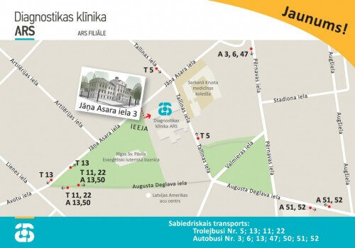
ARS Diagnostic Clinic
Address: J. Asara Street 3, Riga, Latvia
3 Tesla Magnetic Resonance Imaging telephone number:
- Telephone number for appointments: + 371 672 01 088
- Information about the results of the examinations: +371 66 929 760


