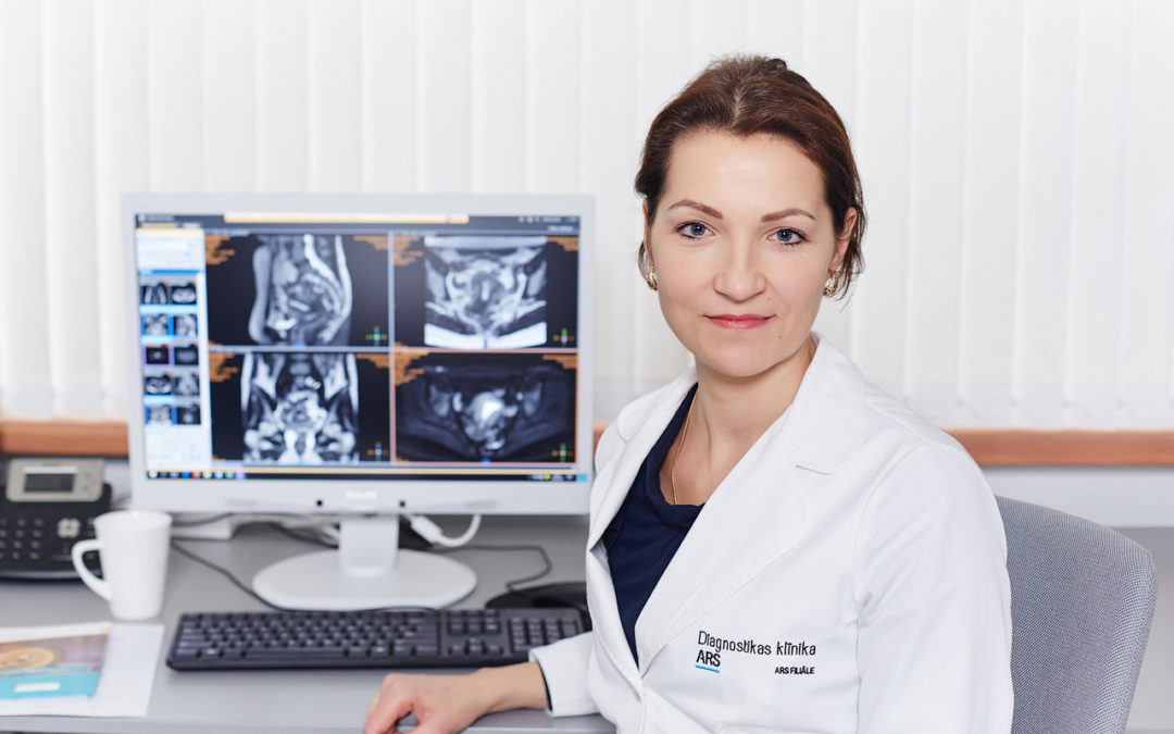
Examination of the lesser pelvic organs is performed at the ARS Medical Centre and its branch, ARS Diagnostics Clinic, using the latest Philips Ingenia 1.5T and 3T systems. Dr Evija OLMANE, Diagnostic Radiologist, M.D., explains the procedure.
Female and male lesser pelvis MRI exams:
Women: gynaecological, bladder, rectum, pelvic lymph node examination
Men: bladder, rectum, pelvic lymph node and prostate examination
When is an MRI exam necessary?
An MRI exam is an informative examination with excellent soft tissue resolution. An MRI exam is performed to determine precisely the pathology and its local spread, i.e. whether or not the disease has spread outside the specific organ system. An MRI exam is performed in case of suspicion of cancer.
A diagnosis is most often based on an oncological disease. An MRI exam is performed to determine precisely the spread of the tumour specifically at the pelvic level in order to select the most appropriate method of therapy, assess the efficiency of the treatment and control the course of the disease. It is therefore very important for a patient to be referred for an MRI exam by a medical specialist, a gynaecologist, urologist or oncologist, who is familiar with the patient’s medical history and his or her current health condition. This is very important information that helps make as precise a conclusion as possible based on the results obtained in an MRI exam.
MRI exams help assess the pelvic organs in case of:
- endometriosis,
- uterine fibroids,
- adenomyosis,
- cervical cancer,
- ovarian cancer,
- prostate cancer,
- colon cancer,
- bladder cancer,
- chronic pelvic pain of unclear origin,
- increased pelvic lymph nodes,
- tumour postoperative control.
Is the more powerful 3T MRI system better?
It is assumed in the world, including Europe and North America, that a 1.5T MRI system is completely sufficient to perform these exams. However, where it is possible to use both systems and to compare the image quality, one must admit that images obtained in a 3T MRI scan have a much higher resolution which facilitates diagnostics greatly.
Preparing for an MRI scan
In order to obtain high-quality images the patient must make certain preparations in advance:
- Fasting (going without food) is required. You should not eat anything at least 4 hours before the procedure.
- You should take the results of all previous medical exams, in-patient or out-patient discharge records, test results etc. related to the specific disease with you. A summary of information on what has happened to the patient prior to the MRI exam should be provided.
The duration of an MRI scan is 20-30 minutes only. It is completely painless. In order to obtain high-quality images, the patient has to lie still without moving during the MRI scan. A set of headphones is provided for the patient in order to reduce the noises made by the machine during the scan.
Complexly harmless
An MRI scan is completely harmless for health because no ionising radiation is used. Tissue resolution is very good, therefore administration of an intravenous contrast agent is required in rare cases only.
For your information:
MRI exam results are available in a digital format and may be forwarded to physicians in other medical clinics.
ARS Diagnostics Clinic,
a branch of the ARS Medical Centre,
Address: 3 Jāņa Asara Street, Riga, Latvia
3T MRI Phones:
- To make an appointment please call (+371) 672 01 088
- For information about MRI exam results please call (+371) 66 929 760


