
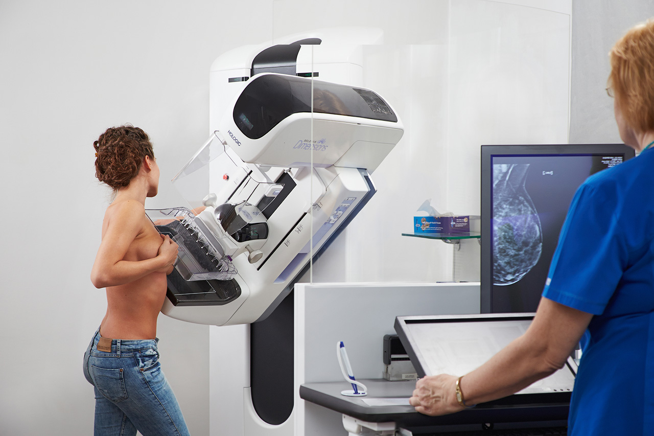
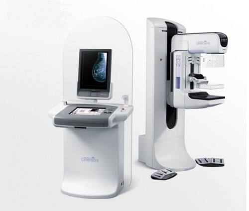
Early breast cancer diagnostics with the digital mammography equipment Hologic Selenia Dimensions, supported by the unique tomosynthesis technology.
New technologies open up new opportunities
The development of science and medicine technologies offer ever more advanced examination methods to diagnose breast cancer at as early a stage as possible. The most popular and most informative method in breast cancer diagnostics is still mammography.

Radiologist-diagnostician Dr. Biruta Rasma VAGULE
Radiologist-diagnostician Dr. Biruta Rasma VAGULE relates:
Unfortunately, breast cancer tends to get increasingly “younger”. In order to reduce the risk of developing a breast cancer, every woman should watch her health, and particularly the health of her breasts throughout her life.
Up until now, as the traditional mammography examinations were conducted, it was very difficult to discover small growths in the mammary gland tissue with dense structure often found in younger women. Because of the limited possibilities of mammography, it was often necessary to supplement it with ultrasound (US) or magnetic resonance imaging (MRI).
In the past few years, digital mammography with tomosynthesis has been used for breast examination in the world. The technology of tomosynthesis is the most effective achievement in the development of mammography within the past 30-40 years. Tomonsynthesis ensures certain diagnostic advantages for women with a mammographically dense breast structure and opens up possibilities for early diagnosis which cannot be achieved with traditional mammography.
Women after 40, should have mammography done once every two years! A timely diagnosis lets cancer to be discovered at a very early stage and it can be successfully treated.
The new mammography equipment Hologic Selenia Dimensions is used for:
- Mammography –2D examination of mammary glands that allows breasts to be examined in different positions. The obtained pictures are digitally processed and stored in an archive, so that, conducting repeated examinations in the future, it is possible to compare and analyze the images.
- Mammography with tomosynthesis – in depth mammary gland examination with the combined method ( for that reason, it is sometimes called the Combo examination), which involves both 3D and 2D examinations. During one examination, actually two are made – digital mammography (2D) and an in-depth examination, using mammography with tomosynthesis (3D). The combined mammography with tomosynthesis is substantially more precise: it allows suspicious changes and possible pathologies in the mammal glands to be discovered much earlier.
 |
 |
How is the examination conducted?
Both 3D and 2D examinations are conducted during a single compression. First, an in-depth examination is done with tomosynthesis; it involves scanning the mammal gland by 1 mm thick layers. The x-ray travels through the breast at a 150 degree angle, taking a series of x-ray images, from which a mathematical 3D reconstruction is made, creating a digital picture that allows the mammary gland to be evaluated in a detailed way. Right after the tomosynthesis, the normal 2D digital mammography is done automatically and, within a few seconds, the digital programme provides the radiologist with a perfectly compatible 2D and 3 D images. Unique imaging technology provides the possibility of obtaining the results in three-dimensional images formed by different projection angles.
F.Y.I.:
Tomosynthesis is an x-ray examination that lets suspicious growths in mammary glands to be examined in great detail.
*Tomos in Greek means ‘slices’.
Advantages of tomosynthesis compared to standard mammography:
- During one examination, both the in-depth 3D and 2D can be done.
- The possibilities of early cancer detection, which would be impossible with standard 2D mammography because of pronounced density of breast tissue.
- Invasive breast cancer detection up by as much as 40 % .
- Primary (initial) detection of all types of breast tumours increased up to 27 %.
- The number of erroneous or the so-called false positive detections decreases by 15%; the impression of a present tumour is caused by the varied appearance of mammary gland tissues.
- During a 3D examination, it is possible to view and make a detailed evaluation of some parts of the breast. That allows for a much more precise examination than 2D.
- The equipment itself records and automatically marks the suspicious structures on the image so that the doctor would pay them closer attention.
- 3D examination reduces the possible need for a repeated mammography.
Pluses of the new Hologic Selenia Dimensions equipment:
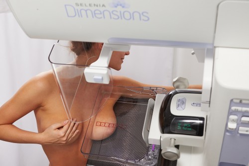
- The examination is painless. It is done with a moveable spatula that adjusts to the shape of the breast. According to the physiology of the woman’s body, the equipment registers the force with which the spatulas of the equipment should be squeezed. The area of the breast area to be examined is sound automatically, including the entire gland to the nipple.
- Comfort has improved and the amount of radiation has been substantially reduced during the examination. For one projection, the examination lasts only 4.5 seconds.
 |
 |
After the examination
The images obtained during mammography are evaluated by the doctor-radiologist who delivers a detailed description and a conclusion. This can be obtained within one week but in emergency cases, immediately after the examination.
F.Y.I.: the results of diagnostic mammography are evaluated by one radiologist but the results of the mammography screening paid for by the state are always examined by two radiologists.
Prices:
- With a specialist’s referral
- The examination is covered by insurance (depending on the type of policy).
ARS Diagnostic Clinic
Address: Jāņa Asara iela 3, Riga
Mammography and Mammography with tomosynthesis:
- registration for the survey: +371 672 01 007
- information about the results of the survey: +371 66 929 750
The mammography results are evaluated by doctors-radiologists:
