
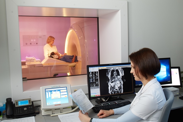
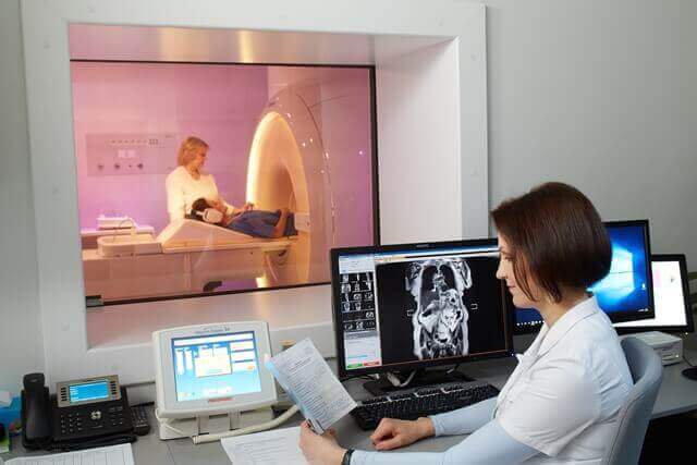
ARS Diagnostic clinic, part of Medical Company ARS, carries out high-resolution examinations with the only newest generation magnetic resonance digital equipment in the Baltics, the Philips Ingenia 3,0T. This equipment facilitates organ diagnosis and therapy in the abdominal cavity and pelvis
In what cases is it necessary to undergo an MRI exam of abdominal or pelvic organs?
- Abdominal cavity exams are intended for the liver, bile duct, gallbladder, pancreas, spleen, adrenal gland, kidney, lymph node, large blood vessel and partly gastrointestinal tract.
- Pelvic exams are intended for the bladder, lymph node and rectum. Also for uterus and ovary examinations in women and prostate examinations in men.
- MRI angiography provides information on vascular pathology – aneurysm, malformation and stenosis.
- MRI provides information about abdominal front wall – subcutaneous formations or hernias.
- Examination can be performed if the diagnosis from previous exams -such as ultrasonoscopy, computed tomography, endoscopy – is ambiguous.
- This examination can be used to establish the spread of previously diagnosed diseases, monitor the course of a disease and the effectiveness of therapy.
- Examination can be carried out in-depth by administering a contrast agent intravenously.
Magnetic resonance imaging provides an opportunity to perform non-invasive examination of the organs of abdominal cavity and pelvis in order to diagnose any pathologies. MRI examination uses a magnetic field and radio-frequency signals to, with the help of a computer, produce high-quality radiological images. These help to assess even the smallest structures in detail.
Abdominal and / or pelvic organ examination with the Philips Ingenia 3,0T MRI is recommended for following impairments:
-
Diffuse hepatic changes
- hepatic steatosis, haemochromatosis, cirrhosis or cardiovascular disease, benign and malignant liver formations.
-
Gallbladder diseases
- cholecystitis, gallstones and gallbladder formations.
-
Biliary tract diseases
- cholangitis, bile duct stricture and bile duct stones.
-
Pancreatic disease
- pancreatitis, benign and malignant pancreatic formations.
-
Spleen diseases
- ambiguous growths.
-
Lymph node pathologies
- lymphoproliferative disease and metastasis.
-
Small bowel diseases
- inflammation, such as Crohn’s disease, benign and malignant formations.
-
Gastric diseases
- benign or malignant formations. Examinations are meant to be performed in order to determine the spread of a disease (examinations are not intended to disprove the gastric inflammatory or oncological existence).
-
Colon diseases
- inflammations (fistulae and abscesses), malignant and benign growth diagnosis, prevalence of the disease and therapy monitoring.
-
Front abdominal wall abnormalities
- subcutaneous formations and abdominal wall hernias.
-
Bladder diseases
- benign and malignant formations, spread of disease and monitoring of disease.
-
Prostate diseases
- hyperplasia, inflammation, malignancy diagnostics, prevalence determination and monitoring of disease.
-
Gynaecological diseases
- congenital abnormalities, inflammations, endometriosis, benign and malignant diagnosis, prevalence determination and monitoring of disease.
How to prepare for an MRI examination:
- No food 4-6 hours beforehand.
- Patients are allowed to drink still water. If a patient comes in for a pelvic organ examination their bladder must be full.
- To maximize the use of MRI diagnostic capacity, patients need to bring all previous documents relating to the disease in question. This includes ultrasound, computed tomography, endoscopy results and statements of previous surgery, manipulations or other treatments, extracts from the hospital, etc.
- The patient’s blood must be analysed to determine their creatinine levels, the results must be taken to the MRI examination.
MRI should not be performed if a patient has:
- a pacemaker,
- nerve stimulators,
- cochlear implants,
- heart valve prostheses or various other metal implants specified as unsuitable for MRI (information will be provided by the attending physician who will refer the patient to the MRI exam),
- metal foreign bodies or claustrophobia (fear of enclosed space).
Useful information:
If an in-depth examination, with a contrast agent administered intravenously, is needed, the radiologist will inform the patient. In order to ensure maximum safety, patients must inform their radiologist of any kidney diseases, allergic reactions and medications used at the time of the examination.
Important:
if a patient suffers from claustrophobia – fear from enclosed spaces, the attending physician can administer calming medication prior the procedure.
Examinations last 20-40 minutes.
Good news:
Philips Ingenia 3,0T is the only MRI equipment in Latvia with a 70 cm wide gantry – cylinder aperture, into which the body part in question is positioned. A wide aperture is well suited for examining larger patients (up to 150 kg ), it also facilitates examination for patients who experience restricted mobility due to pain. This equipment can be adjusted to make the patient comfortable, so that they do not have to endure pain while exam is taking place. This feature helps the patient to maintain a resting position that guarantees better picture quality.
Where it takes place:
MRI examinations of abdominal cavity and small pelvis organs are carried out at Medical company ARS branch –
ARS Diagnostic clinic:
Address – Jāņa Asara ielā 3, Rīgā,
Magnetic Resonance Imaging (3 tesla):
- registration for the survey: +371 672 01 088
- information about the results of the survey: +371 66 929 760