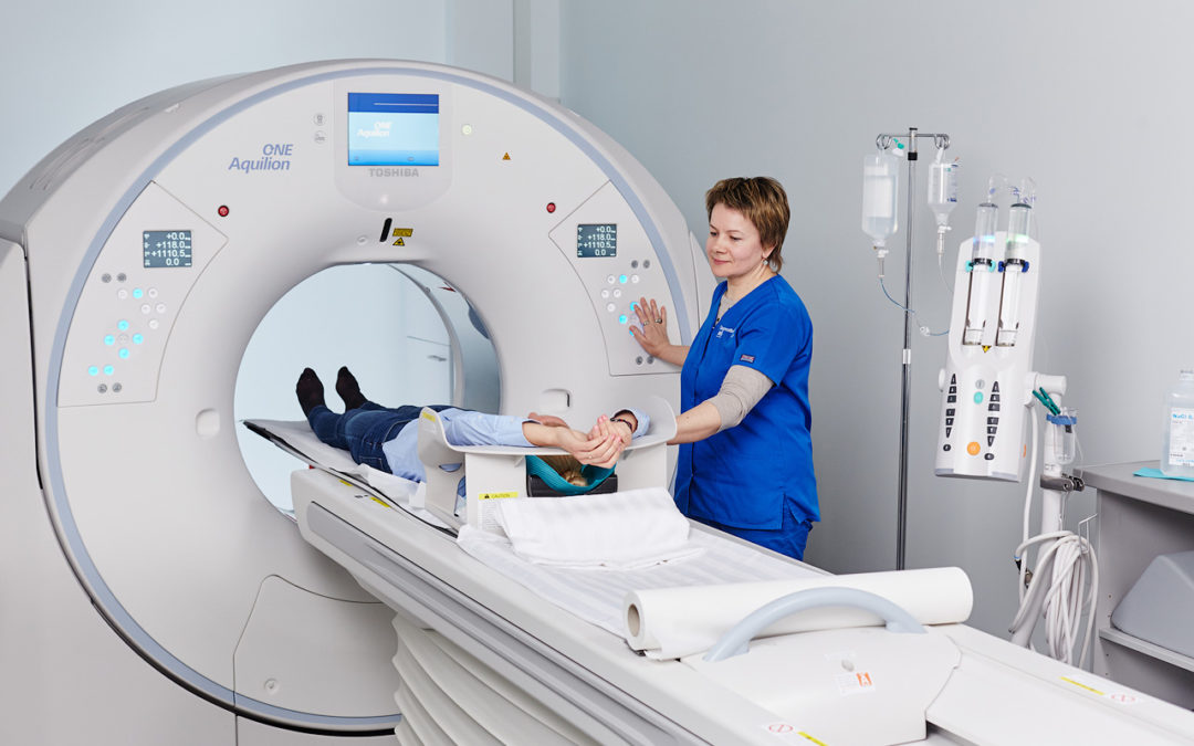
At ARS Diagnostic Clinic in spring 2017 a state-of-the-art Toshiba Aquilion One 320–slice computed tomography equipment was installed.
A new computed tomography scanner Toshiba Aquilion One, developed by a Japan medical devices manufacturer Toshiba Medical Systems Corporation, currently is the only one in Baltic Region. In the nearest region there are two CT devices in Scandinavia – at Karolinska University Hospital in Sweden and at Oslo Clinic in Norway.
Radiologists – diagnosticians of the Medical Center ARS, by using innovative technologies of Toshiba Aquilion One 320 – slice CT, provide computed tomography diagnostic opportunities at a new level.
Advantages of new equipment:
- In comparison with standard CT equipment, the new technology ensures much wider band scanning, thereby significantly reduces the examination time, radiation dose received and improves the quality of images obtained.
- Significantly improved diagnostic opportunities: the obtained data enable to perform an unlimited number of reconstructions with various parameters, depending on the diagnosis and doctor’s requirements.
- Unique gantry opening enables to perform examination changing the angle within the range +/-30°to avoid radiation exposure of sensitive organs.
- Technology of the state-of-the-art detectors ensures basic examinations with a considerably lower radiation dose (up to 40%), as well as additional various screening examinations. E.g., when performing lung examination with an extremely low radiation dose, the patient receives the same radiation dose as through X-ray device, but the diagnostic opportunities are considerably higher.
- Short rotation time of the detector and lamp system (just 0.275 sec – 360° revolution) enables to perform up until now impossible examination of various joints in motion, as well as complex cardiac examinations.
Important information:
Toshiba Aquilion One 320 – slice CT scanner
- Large and comfortable patient table – 47cm wide.
- Gantry opening 78 cm, – now suitable also for tall patients
- The maximum bearing capacity of the table is 220 kg
- The maximum scanning length is 200 cm
- Possibility to adjust the examination angle ±30°
- Unique laser positioning system
- Reduced X-ray radiation exposure
- Shorter examination time
- Wide choice of the examination programmes
New programmes ensure CT examinations:
-
Head
- paranasal sinuses
- skull bones
- brain
- middle ear, mastoid processes
- eye orbits
-
Neck soft tissues
-
Spine, extremities and joints
-
Thoracic organs:
- lungs
- chest wall
- mediastinum, pleural cavity
-
Lungs screening examinations
-
Abdominal organs and small pelvis:
- Liver, gallbladder, bile ducts
- pancreas
- spleen
- adrenal glands, kidneys, ureter, urinary bladder
- lymph nodes
- possibility to gain information also on the gastrointestinal tract, vertebral column and pelvic bones, muscles, pelvic organs, anterior abdominal wall etc.
-
Heart and coronary blood vessels – CT coronary angiography
-
Large blood vessels – CT angiography
Important!
Contrast agents are used in CT examinations to improve diagnostic opportunities.
Opinions of 3 radiologists – diagnosticians:
MD Zanda KRASTIŅA
specialised in thoracic, abdominal and cardiovascular radiology
Examination of the heart and coronary blood vessels using CT equipment significantly reduces X-ray radiation exposure and the examination time. Moreover, the application of the radiation reduction programmes significantly reduces the radiation dose received during examination. Advantage: now it is possible to examine the heart within one heartbeat, if the patient is prepared in advance, by using medication and if the heart rate is about 50-55 beats per minute. Within one heartbeat it is possible to get complete information of the cardiac cavity, myocardium, valves and coronary arteries anatomy, abnormality and functions.
MD Ilze PRIEDĪTE
specialised in thoracic and cardiovascular radiology
Thoracic examinations with new CT equipment can be performed in a shorter time. This significantly reduces the patient breath holding time and facilitates the examination procedure, as well as reduces the contrast agent quantity. The radiation reduction programmes reduces radiation received. The aforesaid is very important for oncological patients and in case of chronic pulmonary diseases, in performance of follow-up examinations. Furthermore, also low radiation doses are possible in the lung parenchyma CT screening examination for the identification of small pulmonary nodules, the radiation dose is similar to X-ray. It is possible to perform the lung and abdomen combined CT examinations – in a single session, the foregoing reduces the radiation dose and contrast agent quantity, which is important for oncological patients, both in the initial examinations for the evaluation of spaciousness of the disease process and in follow-up examinations after therapy.
MD Evija OLMANE
specialised in abdominal radiology
In examination of abdominal organs, new CT equipment renders a number of important advantages: considerably reduced X-ray radiation dose. The amount of intravenous contrast agent to be administered is calculated automatically according to the patient’s weight, thereby reducing the dose of the contrast agent. The examination time is short.
A special programme with extremely low X-ray radiation dose is available, the foregoing is specifically important for oncological patients, when CT examinations are performed repeatedly.
Examinations with Toshiba Aquilion One 320–slice CT are performed by radiologists – diagnosticians:
- MD Zanda KRASTIŅA – specialised in thoracic, abdominal and cardiovascular radiology
- MD Ligita ZVAIGZNE – specialised in thoracic, abdominal and cardiovascular radiology
- MD Evija OLMANE – specialised in abdominal radiology
- MD Ilze PRIEDĪTE – specialised in thoracic and cardiovascular radiology
- MD Helmuts KIDIKAS – specialised in vascular and invasive radiology
ARS Diagnostikas klīnika
3 Jāņa Asara str., Riga
Ph. (+371) 67201007